

Revolutionized is reader-supported. When you buy through links on our site, we may earn an affiliate commision. Learn more here.
Magnetic Resonance Imaging (MRI) is a powerful diagnostic tool that provides highly detailed images of internal structures without ionizing radiation. Unlike X-rays or CT scans, which rely on density differences, MRI works by manipulating the magnetic properties of hydrogen atoms within the body. This technology exploits nuclear magnetic resonance (NMR) principles, using strong magnetic fields and radiofrequency (RF) pulses to produce images with exceptional contrast and resolution. How does an MRI work?
Explore their intracicies, including the underlying physics, image formation, hardware components and the role of artificial intelligence (AI) in advancing MRI technology.
At the core of MRI is the behavior of hydrogen atoms, which are abundant in human tissues due to the high water content. Each hydrogen nucleus behaves like a tiny magnet because of its intrinsic property called “spin.”
When placed inside a strong magnetic field, protons orient themselves either in the same direction as the field or the opposite, generating a net magnetization in the body. Here’s how MRI uses magnetic fields and radio waves:
MRI data is collected in a mathematical space called “k-space,” where signals correspond to different spatial frequencies rather than direct images. Transforming this data into a meaningful image requires sophisticated reconstruction techniques. Below are the key steps in image formation:
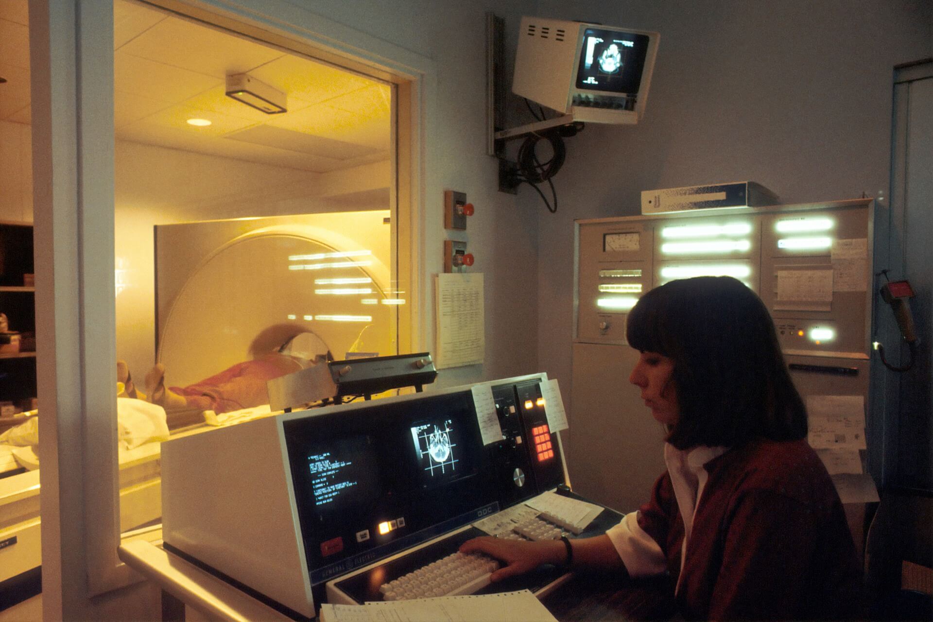
An MRI scanner is a complex system composed of several key components that work in concert to produce high-resolution images.
This is the primary component that creates the strong and stable magnetic field. It consists of coils cooled with liquid helium to maintain superconductivity, ensuring efficient magnetic field generation.
These smaller electromagnets introduce slight variations in the magnetic field, allowing for spatial encoding of signals. Gradient strength determines resolution and scan speed.
These act as both transmitters and receivers. They send RF pulses to excite hydrogen protons and then detect the emitted signals. Advanced coil designs, such as phased-array coils, improve signal sensitivity and image quality.
To prevent electromagnetic interference, MRI scanners are housed in RF-shielded rooms, ensuring that external signals do not corrupt the imaging process.
MRI has evolved beyond simple anatomical imaging. Here are some advanced MRI techniques that provide functional and biochemical insights:
An MRI and a PET (Positron Emission Tomography) scan differ in both their technology and the type of information they provide. MRI uses strong magnetic fields and radio waves to create detailed images of the body’s internal structures, especially soft tissues, while PET scans use a small amount of radioactive material to measure metabolic activity and provide insight into the function of organs or tissues.
MRI is primarily used for visualizing anatomical details, such as the brain, muscles and organs, and it is particularly effective at detecting structural abnormalities. In contrast, PET scans are useful for detecting disease at the cellular level, such as identifying cancer, monitoring brain function or assessing heart disease. While MRI focuses on form, PET focuses on function, providing complementary information in medical diagnostics.
AI is transforming MRI by improving efficiency, accuracy and accessibility. AI-driven algorithms are revolutionizing multiple aspects of MRI imaging.
AI-based techniques, such as deep learning-driven image reconstruction, significantly reduce scan times by intelligently filling in missing k-space data while maintaining high image quality.
AI can automatically detect and segment abnormalities, such as tumors, aneurysms, or lesions, reducing the workload on radiologists and increasing diagnostic accuracy.
Patient movement is a common issue in MRI, leading to image artifacts. AI algorithms can correct motion distortions, improving scan reliability.
AI-powered super-resolution techniques enable high-quality imaging with lower-strength magnets, making MRI more accessible in cost-sensitive settings.
Despite its advantages, MRI faces several challenges that researchers and engineers continue to address.
Traditional MRI scans can take 30 minutes to one hour, causing discomfort, especially for pediatric and claustrophobic patients. AI-driven acceleration techniques aim to reduce these times without compromising image fidelity.
The strong magnetic fields require strict screening for metallic implants, and RF energy can lead to tissue heating. Advances in hardware and monitoring technologies are improving safety protocols.
MRI machines are expensive to purchase and maintain. Efforts to develop low-field, portable MRI systems aim to make this technology more widely available.
Emerging trends include ultra-high-field MRI for unprecedented resolution, quantum-enhanced imaging for better sensitivity and the continued integration of AI for fully automated diagnostics. The convergence of physics, engineering and computational sciences ensures that MRI will remain at the forefront of medical imaging innovation.
MRI is one of the most sophisticated imaging modalities, blending quantum physics, engineering and AI to deliver exceptional diagnostic insights. As advancements in AI, superconducting materials and computational imaging accelerate, the next generation of MRI systems will push the boundaries of speed, accuracy and accessibility.
From revolutionizing neuroscience to enabling early cancer detection, MRI remains an indispensable tool in modern medicine. Continuous innovations are further shaping its future.
Revolutionized is reader-supported. When you buy through links on our site, we may earn an affiliate commision. Learn more here.


This site uses Akismet to reduce spam. Learn how your comment data is processed.