


Revolutionized is reader-supported. When you buy through links on our site, we may earn an affiliate commision. Learn more here.
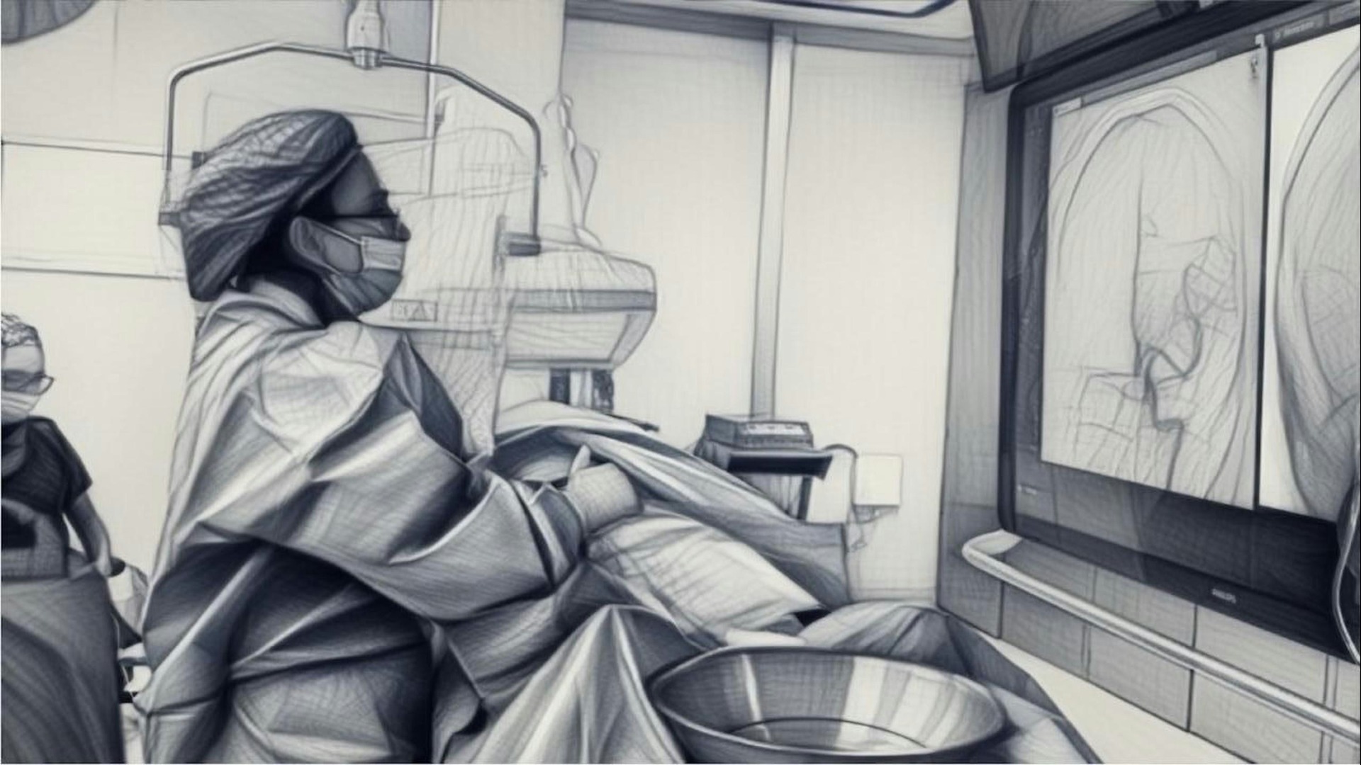
A digital twin is a virtual replica of a physical object, process or person built from real-world data. In health care, surgeons use these doubles to rehearse and refine procedures on a patient’s exact anatomy before even making a single incision. Digital twins in health care are already changing the way operating rooms function, and it will only get better and more precise from here on.
A digital twin built from a patient’s imaging data lets the surgical team test different approaches and instrument paths on a precise 3D model—important for procedures with zero margin for error. Think of it as a full dress rehearsal, where every possible complication is seen on a screen first and not while already in the operating room.
One standout example is Twin-S, a digital twin framework for skull-base surgeries that combines high-precision optical tracking with real-time simulation. During its testing phase, Twin-S achieved an average tracking error of just 1.39 mm while mirroring drilling procedures on anatomical models. The system updates the virtual model at 28 frames per second, giving surgeons augmented views that add an extra layer of situational awareness.
Cardiac surgery is another area seeing major strides. At the Trayanova Lab at Johns Hopkins, researchers build computational replicas of individual patients’ hearts using contrast-enhanced MRI scans. Each digital heart displays scarring, damaged tissue and electrical pathways in a 3D model that the team can rotate and examine from every angle. They simulate heartbeats and apply small electrical signals to pinpoint where disordered rhythms originate, then identify the best ablation sites before the procedure.
Off-the-shelf joint implants do not always match the unique geometry of every patient. A poorly fitting knee or hip replacement can lead to long-term discomfort, limited mobility and even the need for revision surgery. Digital twins address this by creating an exact virtual replica of a patient’s joint and enabling engineers and surgeons to design implants tailored to individual anatomy.
The Hospital for Special Surgery in New York leads this effort. Backed by a $10 million gift, HSS is building a digital twin platform that turns MRI scans into interactive 3D models of a patient’s knee. Surgeons can then simulate how the joint moves and responds to different surgical options. In one early application, the platform enabled surgeons to model ACL revision procedures one degree at a time, predicting how each adjustment would affect knee forces and motion.
A recent study found that orthopedic surgery currently holds the strongest evidence base for digital twin use, with some institutions approaching routine clinical integration. That momentum suggests the industry will likely see these tools become standard practice for joint replacements within the next few years.
MRI and CT scans give doctors flat and cross-sectional images. A digital twin integrates those scans into a dynamic, interactive 3D model that surgeons can rotate, zoom into and peel apart layer by layer. You go from looking at slices on a lightbox to exploring a fully dimensional map of the patient’s internal structure.
This capability is especially valuable in oncology. When a tumor grows close to major blood vessels, nerves, airways or vital organs, knowing the exact spatial relationships makes the difference between a complete removal and leaving cancerous tissue behind.
Researchers are using virtual models and digital twin technology to test new surgical workflows before applying them to patients. Their work focuses on improving safety and quality in cancer interventions through data-intensive modeling.
A review confirmed that patient-specific 3D visualization technologies improve the visibility of neuroanatomical structures at the cranial base. These digital models support approach selection and intraoperative navigation, offering surgeons a far richer understanding of the anatomy than traditional imaging alone.
When powered by biomechanical simulation, a digital twin can predict how living tissue will behave under stress. This is where Finite Element Analysis (FEA) comes in. FEA breaks down a biological structure, such as a bone or a heart valve, into thousands of tiny elements and then calculates how each one reacts to forces like pressure, tension or impact.
Scientists described how digital twins in surgery integrate physics-based modeling with patient data to predict outcomes. For instance, in orthopedic trauma cases, surgeons can simulate how a bone will respond to an implant’s load, or whether a fracture fixation will hold under daily activity. This predictive power lets the surgical team test “what if” scenarios and choose the technique that promises the best biomechanical result for each patient.
Virtual technology is also extending into postoperative care. In a study of over 700 patients, those who underwent both major and minor surgery reported reduced pain while using virtual reality during recovery. As digital twins and VR tools continue to mature, the line between surgical planning and post-surgical support is blurring in ways that benefit the patient from start to finish.
Some digital twins even continue running while an operation is underway. A so-called shadow twin updates the virtual model in real time during surgery, accounting for physical changes, such as tissue deformation and blood loss. Picture a GPS that recalculates the route the moment road conditions change. The shadow twin maps digital information directly onto the patient on the operating table.
Current computer-assisted surgery tools provide limited dynamic, real-time intraoperative information. Digital twins fill that gap by linking the physical world of the operating room with continuously updated virtual data. This is especially useful in minimally invasive and robotic procedures, where the surgeon works through small incisions and relies heavily on visual feedback from screens rather than a direct line of sight.
The Twin-S skull base surgery framework also demonstrates this principle in action. By updating the virtual skull model at frame rate during drilling, it provides augmented views that overlay critical anatomical boundaries in real time. For digital twins in health care, this kind of intraoperative guidance represents one of the most promising frontiers.
Digital twins are making surgery deeply personalized by allowing doctors to rehearse procedures, predict tissue behavior and customize implants for each individual. The focus now turns toward making these models more accessible, so that every patient, regardless of where they receive care, benefits from a surgical plan built especially for them.
Revolutionized is reader-supported. When you buy through links on our site, we may earn an affiliate commision. Learn more here.

This site uses Akismet to reduce spam. Learn how your comment data is processed.