


Revolutionized is reader-supported. When you buy through links on our site, we may earn an affiliate commision. Learn more here.
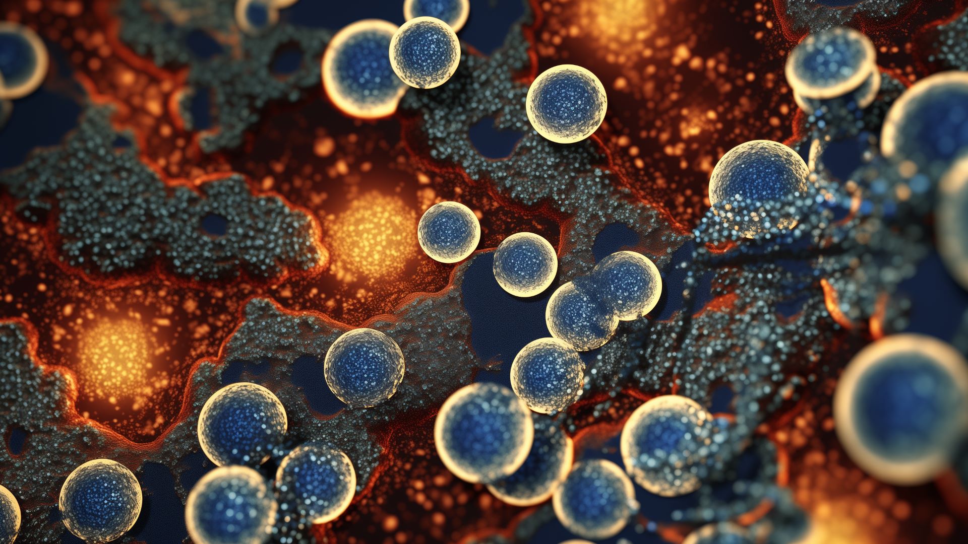
Innovations in biology and technology hinge on understanding the world on a molecular level. The more scientists learn about organisms and materials on the nanoscale, the better they can develop groundbreaking solutions to the world’s problems. Cryo-electron microscopy (cryo-EM) is one of the leading tools enabling that research today.
Cryo-EM has been around for decades, but it’s seen some significant improvements over the past few years. As a result, it’s now one of the best methods at scientists’ disposal for analyzing things on a microscopic scale.
Cryo-electron microscopy is a branch of transmission electron microscopy (TEM) that cryogenically freezes samples before viewing them. It’s become such a significant part of scientific research that it earned three of its developers the Nobel Prize in Chemistry in 2017.
TEM fires beams of electrons at a sample to create digital 3D models of objects too small to analyze with light wavelengths. Cryo-EM takes this further by freezing samples in liquid nitrogen-cooled ethane first. This process suspends any movement in the sample to enable a clear picture and protects it from the electron beam without creating ice crystals that could interfere with the image.
After freezing the sample, scientists use electron beams to generate a few 2D images of it. Specialized software then compares the shadows and combines these 2D images to generate a 3D digital model. Scientists can use that model to analyze microscopic particles or organisms from all sides.
Being able to see tiny samples in detail impacts many branches of research. One of the most popular uses for cryo-electron microscopy is analyzing proteins — the most important compounds in nature. Cryo-EM can reveal protein structures of plants, animal tissues or pathogens to learn how diseases work or make more effective drugs.
Cryo-EM also plays a critical role in bioengineering. Researchers can use it to determine the structures of various spliceosomes or chromosomes. With that knowledge, they can develop novel disease treatments, crop improvements or other helpful gene modifications.
Some researchers use cryo-EM to monitor how neurodegenerative conditions like Alzheimer’s affect human cells or analyze immune system responses. By seeing how these things play out on a molecular level, scientists can learn more about how to prevent some conditions or extend human lives.
Other technologies can help researchers understand microscopic samples, too, but Cryo-EM has some unique advantages. Here are some of the biggest benefits of cryo-electron microscopy.
One of the things that makes cryo-EM so revolutionary is its ability to keep samples in their native state. Conventional TEM can damage organic samples, as the electron beams can be too harsh, or the equipment may require a vacuum. Cryo-EM provides a way around these concerns.
Freezing samples before analysis protects them from damage from the electron beam. Cryo-EM’s use of liquid nitrogen instead of other freezing methods further protects samples. Faster freezing times mean smaller ice crystals, and liquid nitrogen freezes almost instantly, so ice crystals have no time to form.
Instant, crystal-free freezing means researchers can see microscopic samples in their native state, as some properties may change with slower freezing times. That, coupled with the prevention of electron-related damage, makes cryo-EM research more practical than some alternatives.
Cryo-electron microscopy can also use remarkably small sample sizes. That’s because the cryogenic freezing process preserves them in a glass-like state. Other TEM methods often require crystallization, which makes it harder to get a high-resolution image of the sample.
Other TEM technologies compensate for their lack of clarity by using a large concentration of the molecules in question. That way, they can compare and combine a wider range of 2D images to account for any errors or ambiguities that may exist in a few. Those high concentrations can be an issue if researchers are analyzing something particularly rare, expensive or delicate.
By contrast, cryo-EM doesn’t need as high concentrations because each picture of a molecule is clearer from the beginning. As a result, researchers can gain similar insights without needing as many samples of their subject.
The samples that cryo-EM analyzes can be small in more than just their concentration. This technology enables researchers to generate high-resolution 3D models of objects on a near-atomic scale, providing an unprecedented view of some of the world’s smallest particles.
Advances in cryo-EM technology over the past few years have enabled resolutions so detailed that researchers can identify individual proteins inside a cell. Some cryo-EM systems can reveal the positions of individual atoms.
That level of detail could revolutionize advancements in bioengineering, which can lower some medical costs, discover new treatments and enable more functional bionic prosthetics. The more granular scientists can get with their research, the more innovative breakthroughs can result.
The benefits of cryo-electron microscopy are impressive, but it’s not a perfect technology, either. Some challenges remain, making other analysis methods a better choice in some circumstances.
Many cryo-EM scans have issues with noise. One of the reasons this technology can preserve samples’ native state is that it uses lower electron doses on top of freezing the samples. As a result, molecules with low electron absorption like hydrogen and nitrogen — some of the most abundant elements in the universe — can be difficult to render in detail.
Recent advancements have made cryo-EM scans much more detailed. However, noise can still be an issue, especially in older systems without modern upgrades, hindering the analysis of some smaller samples.
Cryo-electron microscopy is also a fairly complex process. There are more steps than conventional TEM methods, and each of these requires high precision. Consequently, the process introduces more room for human error.
Most people using cryo-EM machinery are highly experienced and trained professionals. Still, even leading experts can make mistakes, and even simple errors can jeopardize the reliability of a research project. Automated cryo-EM systems provide a solution, but these technologies are still fairly new and aren’t yet widespread.
Cryo-EM may not be perfect, but it remains one of the best technologies for understanding the microscopic world. As it continues to improve, it could revolutionize the fields of microbiology and biotechnology. Learning about this process, its applications and its benefits is the first step to making the most of it.
Revolutionized is reader-supported. When you buy through links on our site, we may earn an affiliate commision. Learn more here.
This site uses Akismet to reduce spam. Learn how your comment data is processed.