How 3D Brain Imaging Could Help Diagnose Alzheimer’s Disease
February 25, 2023 - Ellie Gabel
Revolutionized is reader-supported. When you buy through links on our site, we may earn an affiliate commision. Learn more here.
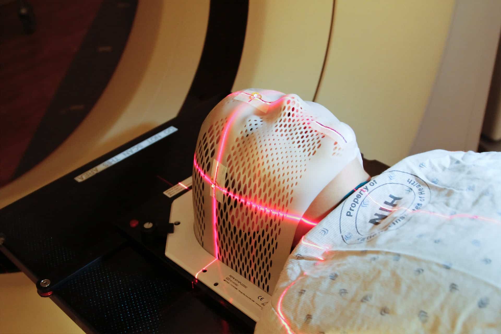
Imagine being able to see tiny blood vessels, lesions and even a person’s pulse through their skull, rotating the image in three dimensions to make a diagnosis and enact a treatment plan. 3D brain imaging is relatively new in the medical field, but it’s one of the most promising forms of technology for people suffering from Alzheimer’s disease. Here’s how recent scientific breakthroughs are shaking up the world of neuroimaging.
Just out of Reach: Diagnosing Alzheimer’s
Alzheimer’s disease is a progressive neurodegenerative condition that affects thinking, memory and behavior. Like sand slipping through an hourglass, the brain gradually deteriorates until the patient loses verbal and motor function.
There is no cure. However, spotting the disorder as early as possible better equips doctors, patients and family members to handle any changes that may occur. A timely diagnosis also allows people to make important decisions regarding life expectancy and care.
Currently, the only conclusive way to diagnose Alzheimer’s disease is through an autopsy. Although positron emission tomography (PET), magnetic resonance imaging (MRI), single-photon emission computed tomography (SPECT) and computed tomography (CT) scans can detect brain shrinkage, misdiagnosis is very common — one study found that 23% of patients diagnosed with Alzheimer’s disease were confirmed not to have it upon autopsy.
Getting the correct diagnosis is crucial. Doctors may otherwise treat patients for Alzheimer’s when they have another condition that requires different care, leading to unnecessary or incorrect medication. That’s where improved 3D brain imaging could be a game-changer.
How 3D Brain Imaging Would Help
Currently, doctors produce 3D brain scans via software that stacks several 2D images — such as from an MRI or PET scan — on top of each other. It allows them to see tumors, lesions, unusual blood flow and other disease indicators. Scientists are always working toward improving current 3D imaging technology, because it could revolutionize the medical field.
One of the main features of Alzheimer’s disease is shrinkage of the hippocampus, a brain region responsible for spatial navigation and memory. Doctors could use new 3D imaging techniques to measure the size of the hippocampus, providing an indicator of disease progression. They could also look for lesions and the buildup of beta-amyloid plaques.
In addition to spotting physical changes, better 3D brain imaging could detect functional differences in the brains of Alzheimer’s patients. For example, functional MRI (fMRI) measures changes in blood flow and neural activity in certain areas of the brain, giving clues as to how the illness affects brain function.
By comparing scans from different points in time, doctors could detect how quickly a patient’s disease is progressing and which areas it is affecting the most. They could then make more accurate prognoses and decide if the patient is a good candidate for experimental drugs or therapies.
Thankfully, there have been several recent breakthroughs in the neuroimaging field.
Untangling a Mystery
A hallmark of Alzheimer’s disease is the accumulation of disordered tau proteins in the brain. As the tau proteins change shape, they form neurofibrillary tangles, clumping up and interfering with cognitive function. What if doctors could identify the tangles as soon as they started to develop?
Last year, researchers at the Karolinska Institutet in Sweden used staining and 3D imaging to detect the accumulation of tau proteins in postmortem human brain tissue, discovering large quantities in the locus coeruleus region. This could give diagnosticians a better understanding of where to look for the first signs of Alzheimer’s disease. It also provides clues as to how the condition starts and spreads within the brain.
Hidden Signals
Another breakthrough is the invention of 3D amplified magnetic resonance imaging (aMRI).
3D aMRI is a new type of 3D brain imaging that detects the movement of blood and cerebrospinal fluid. Within the brain, tiny pulses can indicate things like artery function or blockages caused by a tumor. Traditional MRIs aren’t sensitive enough to pick up on this motion. But 3D aMRI detects and exaggerates the flow of blood and other liquid within the brain, making it stand out to doctors.
In fact, the movements are so amplified — hence the name — that even to the untrained eye, it’s visually obvious that the patient’s brain is pulsing in certain spots. The images are clear and crisp.
This technology is not yet available to the public, but scientists have begun using it in research projects. Coupled with brain mapping to gain a better understanding of how the organ works, 3D aMRI could lead to breakthroughs in diagnosing conditions like Alzheimer’s disease before they become advanced.
Making It Tangible
In 2017, Dr. Darin Okuda used a 3-Tesla 3D brain MRI to scan patients’ brains in remarkable detail. But he didn’t stop there.
After identifying lesions, he uploaded the scans to a 3D printer and made physical models of each damaged area, printing small, white objects that could be mistaken for stones. Holding the lesions in physical form allowed Okuda to examine them with greater precision. His team discovered that lesions from multiple sclerosis (MS) had uniquely identifiable features — they tended to be asymmetrical and had complex surface structures.
In the future, 3D printing may become standard practice in the medical imaging field. Printing out copies of a patient’s brain lesions could help distinguish between Alzheimer’s and other neurodegenerative diseases like MS.
Doctors can 3D print enlarged models to see more detail. Holding a physical model of a lesion, tumor or blocked artery could also make it easier for patients to understand their own diagnoses, especially if they’re having trouble remembering or comprehending spoken language.
The Leading Edge of Neuroscience
Advances in medical imaging are making it easier than ever to detect changes in the brain. That’s great news for Alzheimer’s patients and their families, since doctors will likely be able to use improved 3D brain imaging to detect neurofibrillary tangles, fluid anomalies and lesions before the disease becomes advanced. An earlier diagnosis will allow for prompt treatment and a better care plan.
And, someday, perhaps clinicians can even use 3D brain imaging to find a cure. The future has never looked so bright for patients with neurodegenerative disease.
Revolutionized is reader-supported. When you buy through links on our site, we may earn an affiliate commision. Learn more here.
Author
Ellie Gabel
Ellie Gabel is a science writer specializing in astronomy and environmental science and is the Associate Editor of Revolutionized. Ellie's love of science stems from reading Richard Dawkins books and her favorite science magazines as a child, where she fell in love with the experiments included in each edition.


